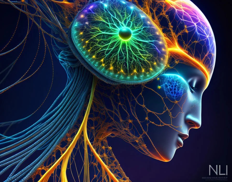
What happens to your brain when your body is asleep
by Marketa Juristova
Sleep is a natural state that is actually the opposite of wakefulness. It is a state in which our physical activity is very much reduced and our consciousness is somehow disconnected from external reality. The body is resting. But what does our brain do when we sleep? Is it also resting?
Like waves in the sea
Let’s go over this from the beginning. You probably know that the brain’s nerve cells are called neurons, and there are an estimated 100 billion of them in the human brain. Didn’t you know that? Never mind, now you know. A neuron consists of its own body, branchlike processes (dendrites) and a central longer process, the axon. Nerve impulses propagate through the axon sheath, like a wave in the sea. These impulses are electrochemically based; it is actually a way of communication between individual neurons. The place where two neurons meet and pass information to each other is called a synapse. So we have a weak electromagnetic field around the brain all the time, which we can measure with an electroencephalogram, or EEG. The way it works is that the person being tested has electrodes attached to the scalp that transmit signals to the EEG, where they are amplified and converted into a graphic record. The EEG recording then shows more or less sharp waves that represent spontaneous electrical activity in the brain. This activity varies, of course, at different levels of mental activity.
If a person is awake, then a large number of so-called beta waves, which are waves with a relatively high frequency but low amplitude (this is simplified to wave height) of 14-30 Hz per second, appear on the EEG recording. When we relax, when the body calms down, when we go to bed, alpha waves appear, which have a lower frequency and a slightly higher amplitude of 8-13 Hz per second. Then, as a rule, comes sleep, which can be divided into five stages, the first four stages not being characterized by rapid eye movements (see previous chapter, NREM).
The first stage is falling asleep, which is the transition between waking and sleeping. In this phase, irregular, low-amplitude theta waves appear on the EEG. If you wake someone up at this time, they will tell you that they have not slept yet.
The second stage, light sleep, is the part where there is a significant reduction in muscle tension. On the EEG, we see larger and slower waves punctuated by so-called sleep spindles (a cluster of waves with a frequency of 15 Hz per second) and K-complexes (large, slow and sharp waves).
This is followed by two stages of deep sleep, deep and even deeper, characterized by delta waves, slow waves with a frequency of 1-2 Hz per second. Here the heart and breathing rates decrease even further and muscle relaxation deepens.
After the deepest sleep stage, when delta waves account for more than 50% of the EEG recording, the person returns to light sleep and then back to deeper sleep. This process repeats for about an hour and a half and then finally, after one of the light sleeps, the fifth stage of sleep, which we call REM sleep, occurs. In addition to the coordinated and rapid movement of the eyelids, the person begins to breathe markedly and irregularly, and the heart rate increases to values normal during wakefulness. The EEG shows similar values to wakefulness, with beta waves predominating along with theta waves.
The first REM phase lasts approximately ten minutes and becomes longer in subsequent cycles, while NREM sleep becomes shorter. During the night, the entire cycle of REM and NREM sleep repeats about five times, waking back to normal reality from the REM phase.
REM sleep is also sometimes called paradoxical sleep, and some scientists consider it more like another state of existence where the body, except for the heart, diaphragm, oculomotor muscles, and smooth muscles, is paralyzed while the brain is highly active.
When we talk about the brain and sleep, we should also mention the special state of consciousness when the body is asleep and the brain is awake, this state is called lucid dreaming. Is this awakening to sleep visible on an EEG?
It has to be said that not much neurological research has been done on lucid dreaming and with a small number of test subjects. This number is in the order of units, rarely tens. This is certainly due to the relatively short time since lucid dreaming has been recognized as an objective condition that can be studied. But the second reason is also the fact that the percentage of people who can lucid dream at least once a week, and would therefore be suitable subjects for study, is still small, which makes it very difficult for scientists both to conduct research and to interpret its results.
In spite of the above problems, there is a suggestion, based on experiments, that this phenomenon is related to the gamma waves (40 Hz per second) that have been measured during lucid dreaming in the lateral (lateral) parts of the frontal cortex, that is, somewhere where we get corners in our hair. But there’s a little hitch. It’s been found that lucid dreaming also involves a higher frequency of eye contractions than the classic REM phase in ordinary dreams. Therefore, the question is whether and how this EEG measurement can be affected by high eye muscle activity. However, gamma waves were also used in another experiment. In 2014, researchers observed a significant increase in lucid dreams when the brain was stimulated with alternating current (tACS) in this frequency band. It was confirmed that the subjects studied actually showed higher self-awareness in the REM phase of sleep after reaching gamma frequencies and felt a separation of their own person from the dream’s plot. Unfortunately, it is somewhat questionable whether these feelings actually meant that the volunteers were experiencing lucid dreams, as there was no objective test method used to confirm lucidity. I’ll get to what that method is in a moment.
Scientific research agrees that lucid dreaming causes higher activation of the autonomic nervous system (e.g. heart and breathing rate, skin potential) than in non-lucid REM sleep. Furthermore, there are at least two neural patterns of lucid dreaming. The first is associated with the „moment of lucidity,“ which is actually a transitory moment when the dreamer becomes aware of his or her own consciousness and is capable of self-reflection. The second pattern captures the „state shift“, the potential persistent differences in brain activity between lucid and non-lucid dreaming in REM sleep.
The brain never sleeps
Imagine that you are alone in a closed room, lying on a mattress that is so comfortable that you can’t even feel it, it is dark, reasonably warm and you can’t hear any sounds from the surroundings. It is just you, alone with yourself. Do you think the brain „shuts down“ in this moment of silence, darkness and stillness?
Which part of the brain is active and how active is determined by its metabolism, and this can be measured well nowadays, for example by positron emission tomography (PET) or magnetic resonance imaging (MRI). Therefore, scientists have already been able to determine which parts of the brain work together in different tasks. We know which area is activated when we observe children playing, a parliament meeting, running up the stairs, solving a maths problem or listening to music. It has been found that when we are left to ourselves without environmental stimuli, high activity appears in areas of the inner frontal cortex, inner and outer parietal cortex, and some other cortical areas. Scientists have called this network (or system) the default or implicit network. Not only my son-in-law, who is an IT specialist, knows that by default/implicit system we mean the default setting. The default system of the brain is actually such a building and functional foundation, a basic setup of many layers and facets of what we call self-awareness, the self.
Think of how often during the day you are thinking about things that have nothing to do with the current environment and situation, planning in your head, talking to yourself. Even in these moments, an implicit system is awakened in your brain that works with some other systems (e.g. visual or auditory cortical fields) like a balance. One system gets dampened and the other gets activated all the more. You know, when you are mentally somewhere else and you are mentally planning, for example, a holiday in Mauritius, you may not immediately notice that a superior has come to your desk because your visual cortical system has just reduced its activity, but the implicit system has started to work more.
As you fall asleep and stay asleep, the map of the active parts of your brain largely coincides with the map of the implicit system. And which parts of the brain are active during sleep? Thanks to magnetic resonance imaging, we know that in deep NREM sleep the brain is slightly more subdued than in conscious rest, but when the brain moves into the REM phase, when a person begins to dream, its activity begins to rise sharply. This happens primarily in the areas where the brain processes visual stimuli, as well as in the motor cortex (the movement centre), in the areas where memory operations are processed, and in the two centres responsible for processing emotions. There, activity increases by as much as 30% compared to the waking state. Conversely, some centres in the lateral parts of the frontal cortex, the so-called prefrontal cortex, which are associated with rational, logical reasoning and decision-making, are deactivated. This means that in the REM phase, centres of the brain related to motor skills, autobiographical memory and emotion processing are active, but the area responsible for rationality and logic ceases to function.
It is thought that the dampening of activity in the prefrontal cortex causes us to believe that even though dreams in the REM phase are so bizarre and nonsensical, we believe them, in the moment, to be reality. At the same time, our self-awareness is lost with the experience of free will. If, despite all this, we realize in a dream that we are actually dreaming, this sleep-deactivated part of our brain suddenly comes back to life.
I mentioned above that gamma frequency testing did not use an objective method to confirm lucidity. This means that there is a way to objectively determine if the person being tested is dreaming or has a false sense of waking up to a dream. Do you know how to tell? It’s in the eyes! They can’t lie.
Give me a signal with your eyes
The term „lucid dreaming“ was coined and used by the Dutch psychiatrist Frederik van Eeden in April 1913 in a lecture to members of the Society for Parapsychological Research. Although a number of popular books dealing with lucid dreaming have appeared in the last century, the subject has remained outside the realm of serious scientific interest.
However, the situation has changed thanks to the American psychologist and lucid dreamer Stephen LaBerge, who began systematic research on lucid dreams in the 1970s as part of his doctoral dissertation at Stanford University.
„Lucid dreaming has been around for thousands of years, but scientific evidence for it has only been emerging since 1975. Graduate student Keith Hearne met Alan Worsley, who described being aware of dreaming in a dream state. Hearne asked himself the question: „How could one prove this?“ Then he had an idea. Hearne noticed that in ordinary dreams there is usually a rapid movement of the eyes. He speculated, „What if I brought this lucid dreamer into a sleep laboratory and asked him to move his eyes from left to right eight times when he became consciously aware that he was dreaming?“ Hearne attached a rapid eye movement sensor to Worsley’s eyes to get a record of them, and placed other sensors on Worsley’s body to make sure he continued to sleep. Alan Worsley became aware that he was dreaming and remembered his task of looking left and right eight times in a row to indicate that he was aware that he was dreaming. He succeeded, providing the first scientific proof of lucid dreaming! He showed that a person can become aware of dreaming even within a dream and signal his consciousness.“ Robert Waggoner
Thus, the confirmation of lucidity by means of eye signals contributed significantly to the recognition of lucid dreaming as a specific state of consciousness by the scientific community, and research on it could begin. In addition, Stanford University also began to call individuals capable of lucid dreaming oneironauts.
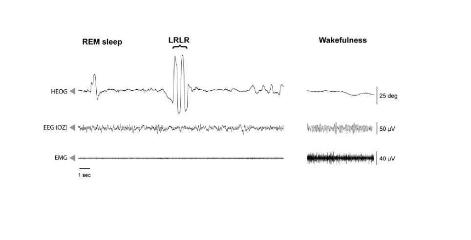
In the figure you can see a sample left-right-left-right-right-center (LRLR) eye movement signal during polysomnographically verified REM sleep. Participants signal that they are aware that they are dreaming by rapidly moving completely to the left (as if to the ear), then completely to the right twice in succession, and then back to the middle without stopping. The LRLR signal can be easily discerned on the electrooculogram (HEOG) output, and has a specific pattern of four consecutive eye movements of a full range of amplitude higher than that of typical REM sleep. The next line shows the high-frequency electroencephalogram (EEG) recording, and below that you can see the minimum amplitude electromyogram (EMG) due to the muscle atonia characteristic of REM sleep (left) compared to the awake state (right). The EMG is a recording of the electrical activity of the muscles. This recording helps in refining the different stages of sleep.
Based on the book by Juristova, M: The White Book of Lucid Dreams -Guide (2021)
Source: BAIRD, Benjamin, Sergio A. MOTA-ROLIM, Martin DRESLER. The cognitive neuroscience of lucid dreaming. Neuroscience & Biobehavioral Reviews. May 2019, 100: 305-323.
Napsat komentář